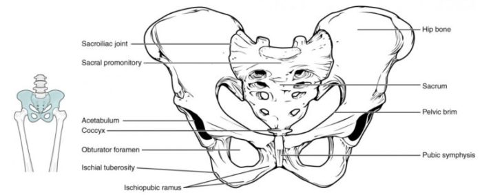
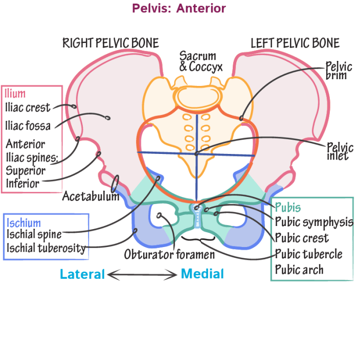
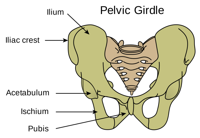
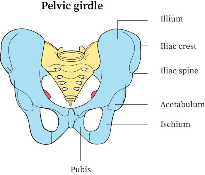
Label the image of the pelvic girdle, an anatomical marvel that forms the foundation of our skeletal framework. Composed of three distinct bones—the ilium, ischium, and pubis—the pelvic girdle plays a pivotal role in supporting our weight, facilitating movement, and providing stability.
Join us as we delve into the intricate details of this remarkable structure, exploring its composition, function, and clinical significance.
This comprehensive guide will unravel the mysteries of the pelvic girdle, providing a thorough understanding of its bony landmarks, articulations, and the intricate network of muscles, ligaments, and nerves that govern its function. We will also shed light on the clinical implications of pelvic girdle injuries, empowering you with knowledge about diagnosis, assessment, and treatment options.
Pelvic Girdle Structure

The pelvic girdle is a complex bony structure that forms the lower portion of the human skeleton. It consists of three pairs of bones: the ilium, ischium, and pubis. These bones are fused together to form a strong and stable ring that supports the weight of the upper body and facilitates movement.
Bony Landmarks and Articulations, Label the image of the pelvic girdle
The ilium is the largest and most superior bone of the pelvic girdle. It has a broad, fan-shaped body that flares laterally to form the iliac crest. The iliac crest is a prominent bony ridge that can be felt at the top of the pelvis.
The ilium also has two prominent projections: the anterior superior iliac spine (ASIS) and the posterior superior iliac spine (PSIS). The ASIS is located at the anterolateral corner of the ilium, while the PSIS is located at the posterolateral corner.The
ischium is a triangular bone that forms the posterolateral portion of the pelvic girdle. It has a body that is fused to the ilium and a ramus that projects inferiorly and anteriorly. The ischial tuberosity is a large, rounded projection at the inferior end of the ischial ramus.
The ischial tuberosity is the primary weight-bearing surface of the pelvis when sitting.The pubis is a small, triangular bone that forms the anteromedial portion of the pelvic girdle. It has a body that is fused to the ilium and ischium and a ramus that projects inferiorly and medially.
The pubic symphysis is a cartilaginous joint that connects the two pubic bones at the midline.The pelvic girdle articulates with the sacrum posteriorly and the femur anteriorly. The sacroiliac joint is a strong, fibrous joint that connects the ilium to the sacrum.
The hip joint is a ball-and-socket joint that connects the femur to the acetabulum, a socket-shaped depression on the lateral aspect of the pelvis.
Parts of the Pelvic Girdle
| Part | Location | Function ||—|—|—|| Ilium | Superior and lateral | Supports the weight of the upper body and provides attachment for muscles || Ischium | Posteroinferior | Supports the weight of the upper body and provides attachment for muscles || Pubis | Anteromedial | Supports the weight of the upper body and provides attachment for muscles || Acetabulum | Lateral | Forms the socket for the hip joint |
Pelvic Girdle Function: Label The Image Of The Pelvic Girdle

The pelvic girdle serves several important functions:*
-*Supports the weight of the body
The pelvic girdle is the primary weight-bearing structure of the lower body. It transfers the weight of the upper body to the legs through the hip joints.
-
-*Facilitates movement
The pelvic girdle provides stability and mobility for the lower body. It allows for a wide range of movements, including flexion, extension, abduction, adduction, and rotation.
-*Protects the pelvic organs
The pelvic girdle helps to protect the pelvic organs, including the bladder, rectum, and reproductive organs.
Pelvic Girdle Anatomy

The pelvic girdle is associated with several major muscles, ligaments, and nerves:Muscles:* Gluteus maximus
- Gluteus medius
- Gluteus minimus
- Hamstrings
- Adductor muscles
- Abductor muscles
Ligaments:* Sacroiliac ligaments
- Iliolumbar ligament
- Inguinal ligament
- Pubofemoral ligament
- Ischiofemoral ligament
Nerves:* Sciatic nerve
- Femoral nerve
- Obturator nerve
- Pudendal nerve
Clinical Significance of the Pelvic Girdle

Injuries to the pelvic girdle can be serious and can have a significant impact on mobility and quality of life. Common pelvic girdle injuries include:* Fractures
- Dislocations
- Ligament sprains
- Muscle strains
Pelvic girdle injuries can be diagnosed using imaging techniques such as X-rays, CT scans, and MRI scans. Treatment for pelvic girdle injuries depends on the severity of the injury and may include:* Rest
- Ice
- Compression
- Elevation
- Medication
- Surgery
Frequently Asked Questions
What are the three bones that make up the pelvic girdle?
The three bones that form the pelvic girdle are the ilium, ischium, and pubis.
What is the primary function of the pelvic girdle?
The pelvic girdle’s primary function is to support the weight of the body, facilitate movement, and provide stability.
What are some common pelvic girdle injuries?
Common pelvic girdle injuries include fractures, dislocations, and ligament sprains.